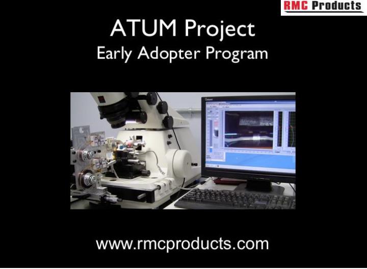
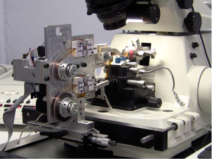
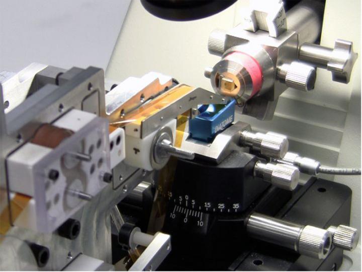
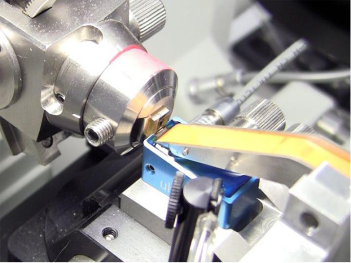
ATUMtome: Automated Tape Collecting Ultramicrotome
The ATUMtome tape collecting ultramicrotome is now available to sophisticated early adopters* for high resolution array tomography of biological tissues.
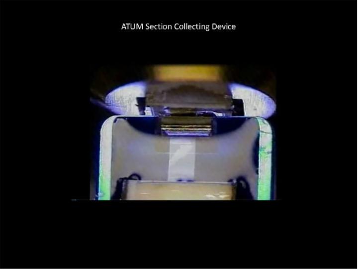
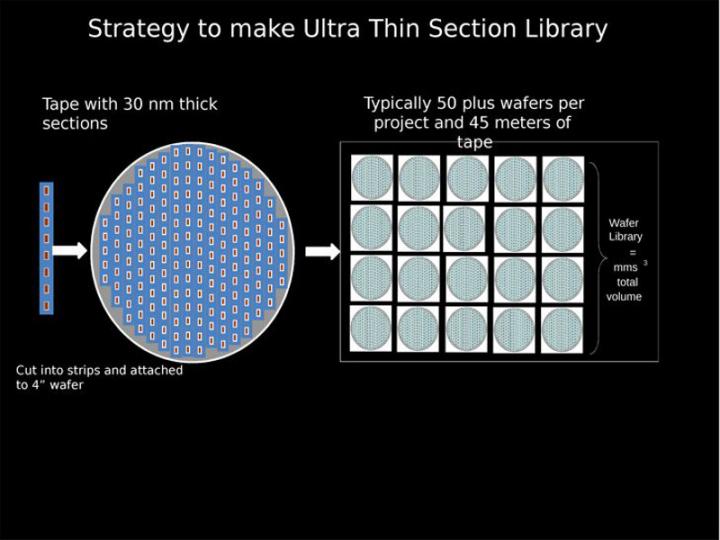
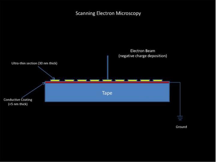
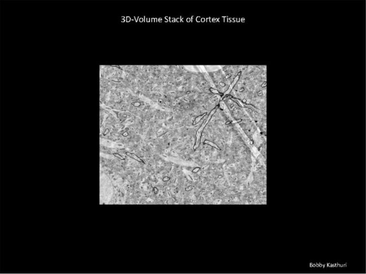
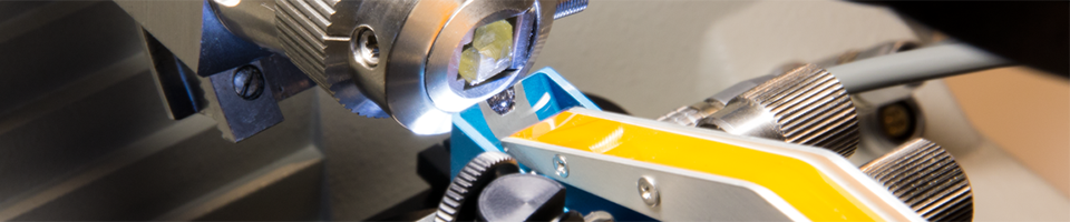
Thousands of utra-thin sections with a thickness of 30nm can be automatically collected on 8mm wide Kapton tape for SEM imaging and sebsequent 3-D reconstruction.
The ATUMtome’s tape collecting part of the system (ATUM) is under license from Harvard University where Professor Jeff Lichtman, PhD, MD, and his team designed the instrument to help collect ultrathin sections to help reconstruct the brain’s neural pathways — a precursor to one day mapping the entire human brain.
* The ATUMtome is available only to sophisticated users during the Early Adopter Program.
Features & Benefits
- Collects hundreds to thousands of sections on a continuous tape
- Non-destructive to sample, with sections available for years into the future for processing, post-staining, immunogold labeling, correlative imaging — and at any work pace desired.
- Section thickness usually range from 30nm to 5,000nm, with thinner sections possible.
- Determines sample viability sooner in the process so you don’t waste valuable time cutting and imaging a problem sample.
- Uses multiple resolutions for locating regions of interest, then zooming in for higher resolution imaging
- Allows correlative microscopy for localization and then ultra-structural imaging
- Prepares samples that allow short pixel dwell time for fast detection of images during electron microscopy
- Uses standard sample preparation techniques and resins
- Charging effect on sections is manageable
- Priced at a fraction of the cost of alternative 3-D imaging techniques
Supplied Complete with:
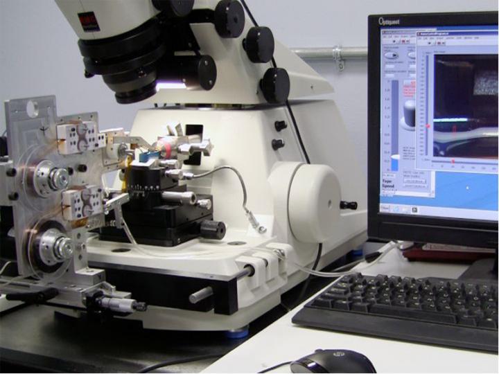
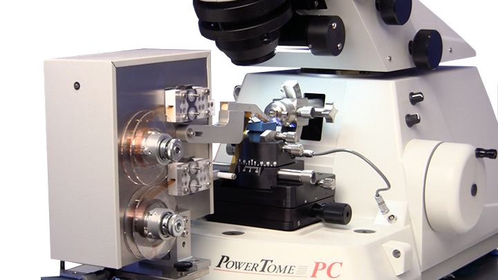
- ATUM continuous tape feed mechanism with PC control software
- PowerTome PT-PCZ
- Air-activated anti-vibration microtomy table including ATUM attachment interface with x-y-z fine control positioning of tape/section pick-up head
- Silent compressor
- Environmental chamber Anti-static device
- Ergonomic lab chair
- 4 mm diamond knife, 35 degrees for room temperature ultra-thin sectioning, mounted in large-cavity blue anodized holder
- Water level control system
- Wafer workstation
- Start-up supply of Kapton tape
- Four 4” diameter silicon wafers
| Dimension: | 49” W x 36” D x 54” H |
| Weight: | 796 lbs. ( 361 kg) |
| Electrical: | input: 110 – 240 VAC 50/60 Hz; output: 255 watts |
Installation Considerations ATUMtome
Pre-Installation Considerations (PDF)
Resources
NEW! ATUMtome Technology Exchange for Early Adopters.
If you are an early adopter, register here to explore techniques, share tips and ideas, obtain the latest news on the ATUMtome and help us to continue developing the product for worldwide research.
ATUMtome News Release – Nov. 7, 2014
Reference Links The Human Connectome Project: Creating a Complete Roadmap of the Brain – VIDEO
Drawing inspiration from the Human Genome Project, neuroscientists in the US want to map all the neural pathways in the human brain, revealing for the first time the physical structure of individual memories and even the faulty wiring that may underlie some psychiatric conditions.
Connectomics: Jeff Lichtman at TEDxCaltech – VIDEO
On January 18, 2013, Caltech hosted TEDxCaltech: The Brain, a forward-looking celebration of humankind’s quest to understand the brain, by exploring the past, present and future of neuroscience. Presenter is Jeff Lichtman, a Jeremy R. Knowles Professor of Molecular and Cellular Biology at Harvard and member of Harvard’s newly established Center for Brain Science.
Automated Serial Sections to Tape by Hall, Hartwieg and Nguyen
Description and operation of the ATUM (Automatic Tape-collecting Ultra-Microtome), which is currently the most advanced means to automate the collection of serial thin sections onto a continuous reel of tape, which can then be stained and viewed by SEM.
ATLAS and ATLAS 3D
Large area imaging for SEM, FE-SEM & FIB-SEM, via Zeiss. Includes applications and downloadable product information.
Automated Transmission-Mode Scanning Electron Microscopy (tSEM) for Large Volume Analysis at Nanoscale Resolution
A research article by Masaaki Kuwajima, John M. Mendenhall, Laurence F. Lindsey, Kristen M. Harris from The University of Texas at Austin, via Plos One
Array Tomography: A New Tool for Imaging the Molecular Architecture and Ultrastructure of Neural Circuits
Published in Neuron, July 2007, by Kristina D. Micheva and Stephen J. Smith. Papers & Other Publications A list of research papers and other publications that mention use of the ATUM.